

Administrator
04 February 2016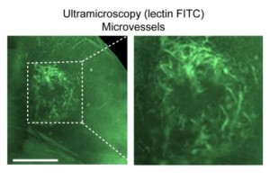
Representational Picture Courtesy : scifeeds.com
Doctors treat brain tumours by attempting to stop the growth of blood vessels in tumours; but imaging methods are essential for initial diagnosis and monitoring the effects of treatments.
Visual mapping of vessels in tumours has also proven a challenge. Researchers have now developed a combined magnetic resonance imaging (MRI) and ultramicroscopy ‘toolkit’ to study vessel growth in glioma models in more detail than previously possible.
Their study is to be published in the journal eLife.
“Gliomas are highly malignant brain tumours with poor prognosis,” says Michael Breckwoldt, a physician-scientist and one of the lead authors of the paper from the University of Heidelberg.
“Many efforts have been made to develop therapies against the growth of blood vessels and therefore ‘starve’ tumours of their resources, but they are not entirely effective,” he points out.
“Improved imaging techniques that faithfully show the vessel architecture, including their growth, structure and density, and the effects of treatments in a non-invasive way are therefore needed to inform the development of future clinical trials.
” In their study in mice, the team combined an MRI approach in vivo (from within) with ultramicroscopy of the whole brain.